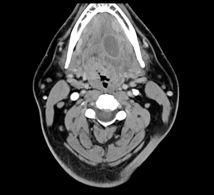
Join Dr. Suresh Mukherji as he explains the ins and outs of the Oral Cavity and Oropharynx. Employ a practical approach to this important and complex topic. In this section, learn the anatomy and various pathologies including neoplasms, infections, and congenital and vascular malformations of this area.
After completing this course, you will be better able to:
- Apply appropriate search patterns to ensure high quality case assessment
- Identify key anatomical landmarks, variations, and abnormalities on imaging
- Accurately interpret advanced imaging cases
- Formulate definitive diagnoses and limited differentials
Faculty and Planning Disclosure
Oral Cavity Imaging: Introduction – 9 min
Left Glossotonsillar Sulcus SCCA CT – 3 min
Right Glossotonsillar Sulcus Ca Classic – 3 min
Floor of Mouth SCCA Carcinoma – 3 min
Left Tonsilar Cancer – 3 min
Alveolar Ridge CA – 3 min
Mandibular Lymphoma – 2 min
ACC of the Oral Cavity – 2 min
SCC of the Tonsil – 3 min
Tonsilar Cancer – Why it’s Not a Glomus Tumor – 3 min
Right Tongue Base SCCA – 3 min
Tonsilar Phlegmon – 2 min
Ludwig’s Angina – 3 min
Oral Abscess – 2 min
Suppurative Adenitis with Retropharyngeal Effusion – 3 min
Pharyngeal Trauma – 2 min
Bilateral Ranulas – 3 min
Venolymphatic Malformation of Right Face/Oral Cavity – 3 min
Lymphatic Malformation – 2 min
Mixed Vascular Malformation – 5 min
Oropharynx Anatomy – 9 min
Course Evaluation


